
Figure 6. Localization of LBs in hippocampal astroglia, microglia and neurons.
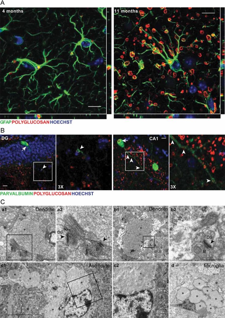
Representative images from 4-month-old (A) and 11-month-old (A–C) malin KO hippocampus.
- Orthogonal confocal sections are shown for astrocytes containing polyglucosan accumulation in the hippocampus of 4- and 11-month-old KO mice. Antibodies were used against GFAP (green) and polyglucosan (red). Hoechst (blue) was used for nuclear staining. Scale bar = 10 µm.
- Confocal images are shown for DG and CA1 hippocampal regions. Antibodies were used against parvalbumin (green) and polyglucosan (red). Hoechst (blue) was used for nuclear staining. Polyglucosan accumulation can be observed within the somas (white arrows) and some processes (white arrowheads) of PV+ interneurons. Scale bar = 10 µm. 3X = 3-fold magnification.
- Electron microscopy images are shown for CA1. Micrographs depict the presence of LBs and glycogen granules in dendrites (a1, a2, b1, b2). LBs were also found in astrocytes (c1, c2). Microglial cells with some engulfed LBs were observed (d). *, Lafora Body; +, glycogen granule; black arrowhead: postsynaptic density; B: synaptic bouton; DS: dendritic spine. a2, b2 and c2 are magnifications of the boxes in a1, b1 and c1. Scale bars are 5 µm in c1 and 0.5 µm in a1, a2, b1, b2, c2 and d.