
Figure 3.
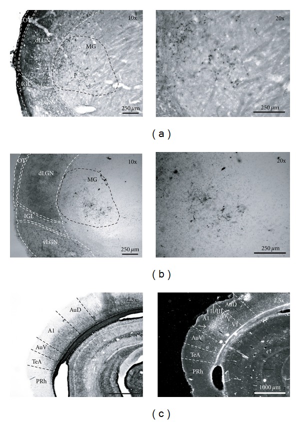
Ectopic retinal projections to the medial geniculate nucleus (MG) in the SC + ICb lesioned (or rewired) hamster. (a) Retinal projections in the MG labelled by intraocular injection of the cholera toxin β fragment and (b) co-tagged with wheat germ agglutinin-horse radish peroxidise (WGA-HRP). (c) Gold chloride myelin staining (left panel) and transneuronal labelling, with WGA-HRP (right panel), of new visual thalamo-cortical afferences reaching namely cortical layer IV in the primary auditory cortex (A1) (white arrow head). A1, primary auditory cortex; AuD, dorsal secondary auditory cortex; AuV, ventral secondary auditory cortex; TeA, temporal association cortex; PRh, perirhinal cortex; dLGN, dorsal lateral geniculate nucleus; vLGN, ventral geniculate nucleus; LP, lateral posterior nucleus; MG, medial geniculate nucleus; ot, optic track; IGL, intergeniculate leaflet.