
Figure 2. Mitochondrial morphology in brains of mice with a conditional knock-out in noradrenergic neurons (DBH-Cre).
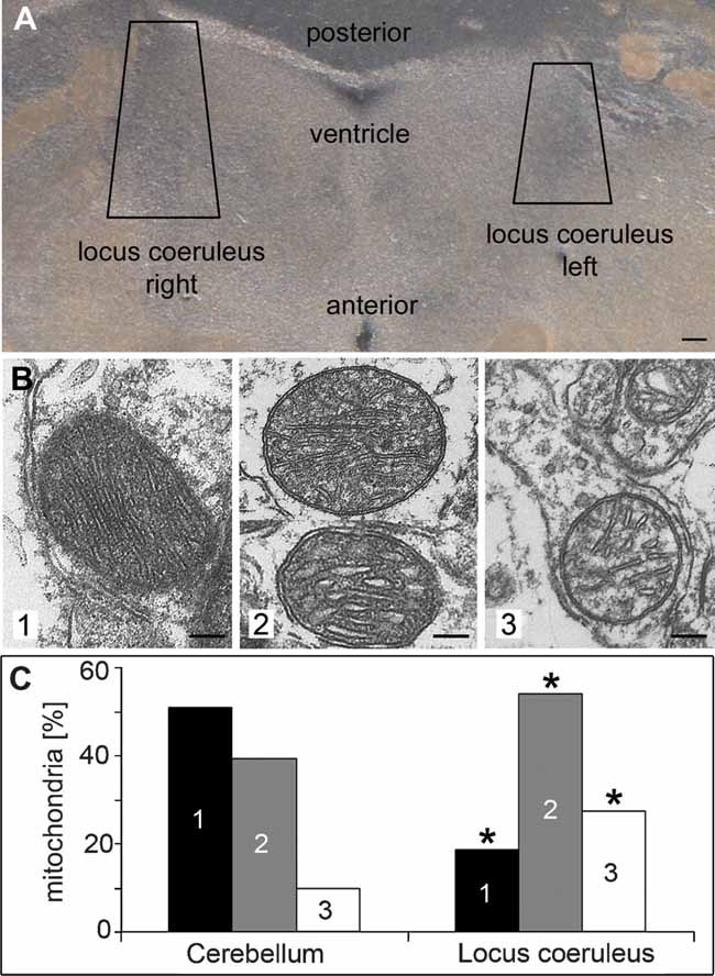
- A. Brains from knock-out mice were Vibratome sectioned (50 µm), the loci coerulei, which could be identified by pigmentation, were dissected and prepared for electron microscopy. Scale bar: 100 µm.
-
B, C. Mitochondria were classified into three groups (1—dense, dark; 2—loosely packed/swollen cristae; 3—depleted cristae). Total numbers per sample and an overview of the cells are given in Supporting Information Fig 5. Sections of the cerebellum, which lacks noradrenergic neurons, served as a control. * Indicates significance (p < 0.0001) of differences to the cerebellum.Pictures of 33 random systematically chosen visual fields were taken in a magnification of 11.5 × 10³, scale bars: 100 nm.