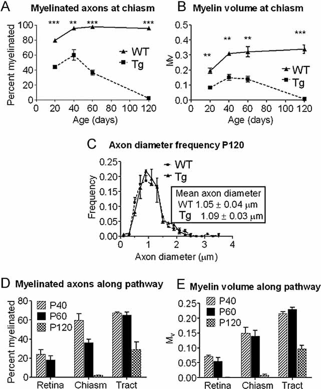
Figure 1. Demyelination progresses in a rostral to caudal direction with increasing age in optic pathway of Plp1-transgenic mice.

- A, B. The percentage of myelinated axons (A) and the relative volume of myelin (B) at the chiasmal end of the optic nerve in wild type (WT) and Plp1-transgenic (Tg) mice between P20 and P120.
- C. Axon diameter frequency distribution at the chiasmal end of the optic nerve of WT and Plp1-transgenic mice aged P120. Axonal diameter distributions are not significantly different.
- D, E. The percentage of myelinated axons (D) and the relative volume of myelin (E) along the optic pathway of Plp1-transgenic mice at P40, P60 and P120 showing the temporal and spatial progression of demyelination.
