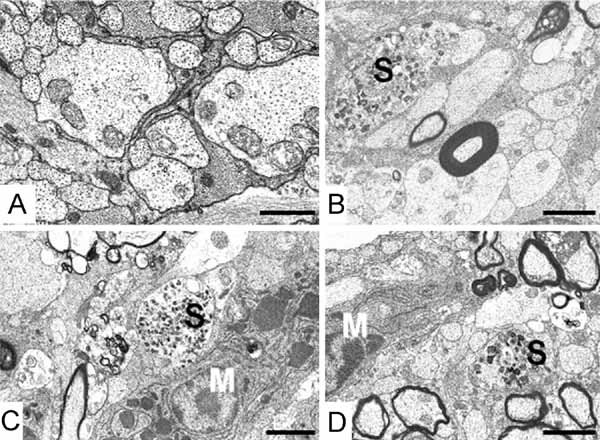
Figure 2. Distribution of myelin and axonal pathology along the optic pathway of Plp1-transgenic mice aged P120.

- A. Retinal end of optic nerve: all myelin has been removed leaving naked axons, which appear morphologically normal and are interspersed with astrocyte processes. Bar: 1 µm.
- B, C. Chiasmal end of optic nerve: demyelination is on-going although most axons have lost their sheaths. Microglia (M) and axonal spheroids (S) are present. Bar: 2 µm.
- D. Optic tract: demyelination is present but not as advanced as at the chiasm. Microglia and axonal spheroids are evident. Bar: 3 µm.
