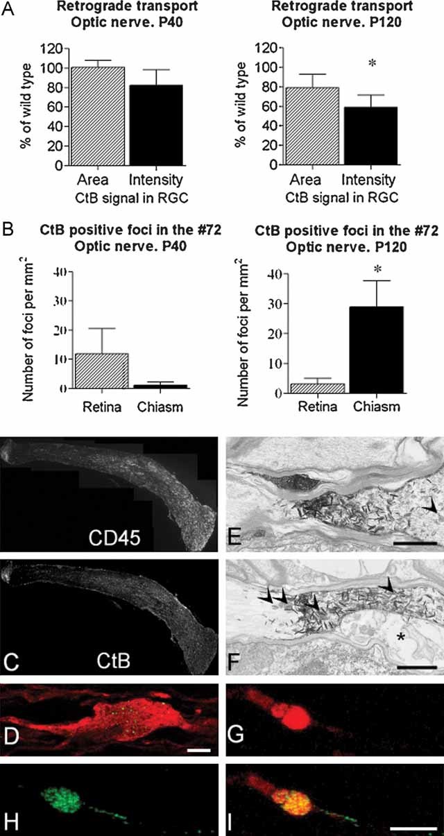
A. Following injection of CtB into the rostral colliculus, the mean signal intensity in the retinal ganglion cells (RGC) and the area of retina occupied by labelled cells was determined in WT and transgenic littermates aged P40 and P120. At P40, there is no significant difference between transgenic and WT. However, the signal intensity is significantly lower in the P120 transgenic group, when there is active, inflammatory demyelination. As the axon counts in the optic nerves of the two genotypes are also similar the data suggest that the reduced signal is due to impaired transport rather than axonal loss.
B. Quantification of CtB foci in retinal or chiasmal portions of the P40 or P120 nerve. At P40, only a few foci are observed and they tend to be more prevalent in the retinal half of the nerve. However, the difference between retinal and chiasmal halves is not significant. At P120, sites of transport stasis are located predominantly in the distal portion of the nerve.
C. Longitudinal sections of nerve (retinal end left) from a Plp1-transgenic mouse aged P120 immunostained for CD45 and showing CtB positive foci. The sections show the gradient in macrophages and CtB between the two ends of the optic nerve. Lipofucsin is present in these nerves, particularly in the chronically demyelinated region, and is indistinguishable from CtB in these images.
D. Single z step through an SMI32-labelled (red) axon in which FITC–CtB (green) appears punctuate, suggesting that it may associate with endosomes and multivesicular bodies. Bar: D = 10 µm.
E, F. Electronmicrographs of horseradish peroxidase (HRP)-conjugated CtB, labelled with TMB, in axons in the optic tract of a P120 mouse. TMB appears as electron dense ‘spikes’. The axons remain patent. Axons in these images remain myelinated, however in (F), the inner tongue (asterisk) of the oligodendrocyte is swollen. Arrowheads indicate axonal organelles. Bar: E,F = 1 µm.
G, H, I. Eighteen hours after TRITC–CtB (red) and FITC–CtB (green) were simultaneously injected into the eye and the rostral colliculus, respectively, confocal microscopy showed that many axonal swellings contained both tracers. This suggests that the swellings are associated with defects in both retrograde and anterograde axonal transport, and also indicates that the associated axon remains intact. Bar: I = 5 µm.

