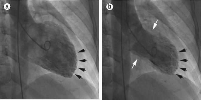
Figure 2.
Left ventriculogram in (a) diastole and (b) systole from the patient with exogenous epinephrine–induced takotsubo cardiomyopathy. The apical segments show essentially no movement (black arrows) relative to the basal segments (white arrows), reproducing the classic “ballooning” that looks like a Japanese “takotsubo,” or octopus trap.

