Figure 3.
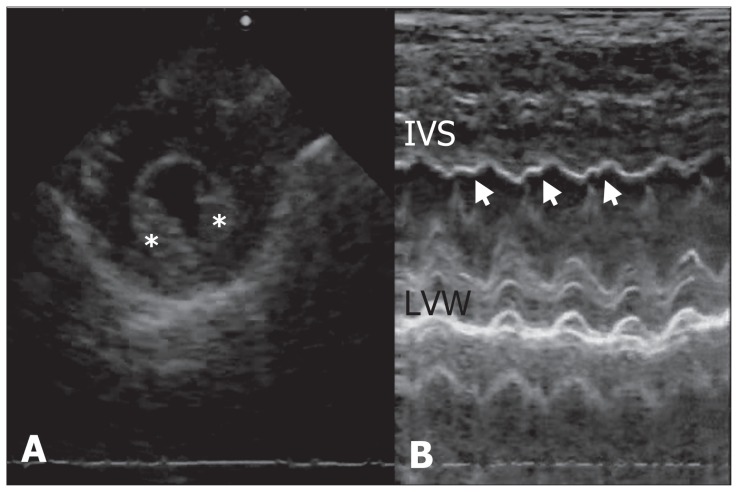
Echocardiograms from a dog with pseudoephedrine toxicosis. (A) M-mode echocardiogram at the level of the left ventricle, showing the hyperechogenic left ventricular endocardium (asterisks). The endocardium was brighter than the underlying myocardium. (B) Abnormal interventricular septal motion was demonstrated on left ventricular M-mode (arrows). IVS — interventricular septum, LVW — left ventricular wall.

