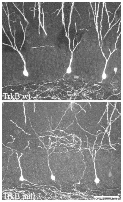
FIGURE 3.
GFP fills both the dendritic and axonal arbors of dentate granule cells in TrkB+/+ and TrkB−/− mice crossed to Thy1 GFP-expressing mice. Images are confocal projections showing GFP-expressing dentate granule cells. All three cells in each image are located along the hilar border, and exhibit a single radially projecting dendrite as is typical for cells located in this region. Some dendrites are truncated at the surface of the tissue section in these images. Scale bar = 50 μm.

