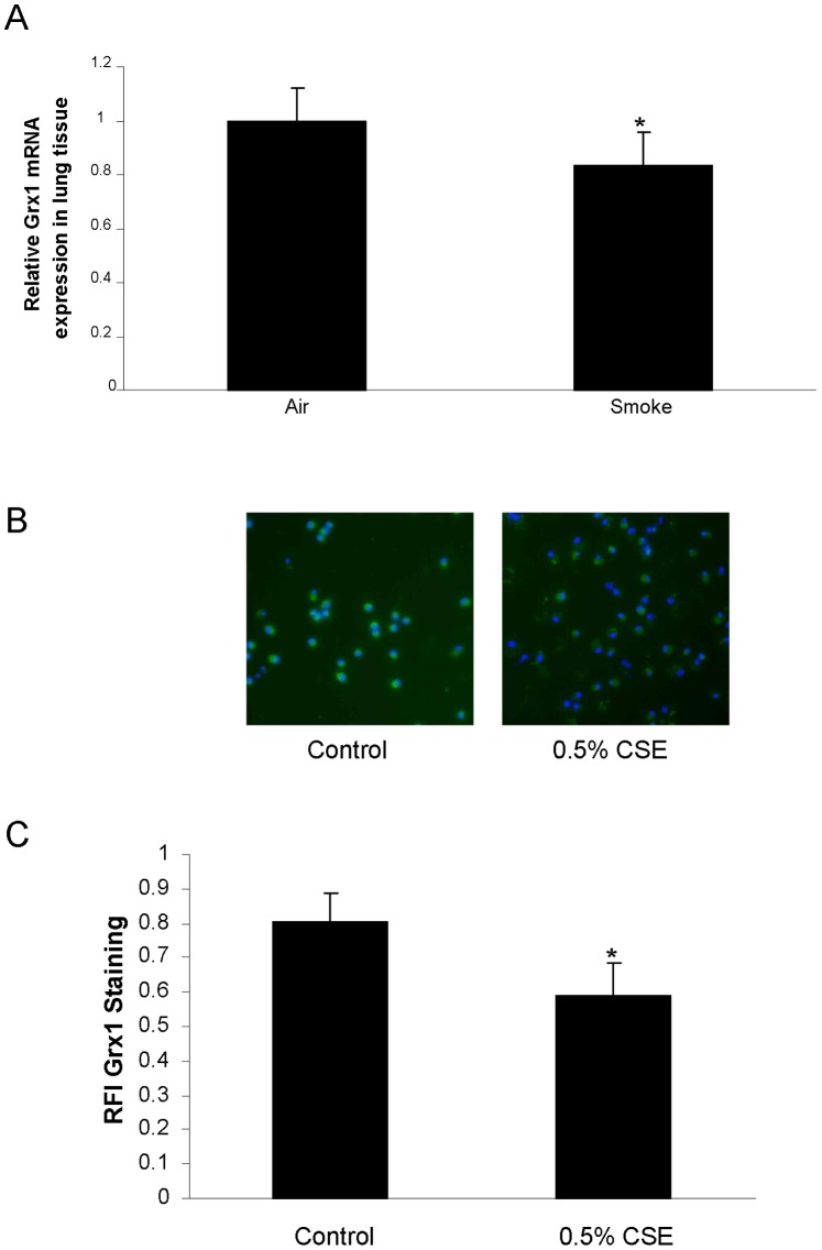
Figure 5. Grx1 expression in lung tissue and primary macrophages of mice exposed to cigarette smoke.
(A) Grx1 mRNA expression corrected for HPRT mRNA expression in lung tissue of mice exposed to air and cigarette smoke, represented as mean ± SD. (B) Fluorescent Grx1 staining in primary macrophages after 24 hours of control or 0.5% cigarette smoke extract exposure. The green staining represents Grx1 protein expression, whereas blue represents the nuclear DAPI staining. Quantification of fluorescent Grx1 staining is expressed as RFI in (C).

