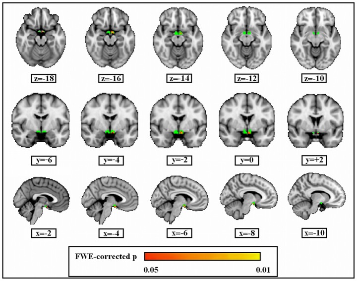
Figure 3. Grey matter volume loss of left lateral hypothalamus in ED patients than healthy subjects.
Green colour describes the extent of the hypothalamus ROI. The significant voxels (family-wise error rate is controlled) are projected onto a Montreal Neurological Institute (MNI152) template and identified by colours ranging from red to yellow (the colored bar represents the p-value). All images are oriented according to radiological convention (the left hemisphere of the brain corresponds to the right side of the image).

