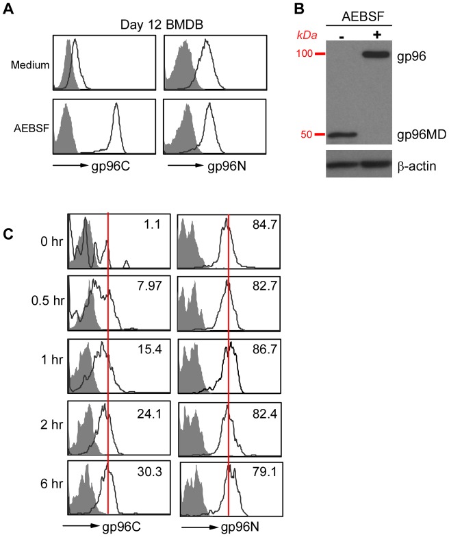
Figure 7. Expression of gp96MD is mediated by proteolysis.
A, BMDBs were treated with AEBSF for 6 hours followed by intracellular stain for gp96 using either C-terminal or N-terminal specific antibody. B, BMDBs were treated with AEBSF for 6 hours followed by subjecting total cell lysate to electrophoresis and immunoblot for gp96. C, Kinetic emergence of intracellular gp96 C-terminal antibody reactivity after treatment of B220−IgG+ population with AEBSF for the indicated time. Number in the quadrant indicates mean fluorescence intensity of gp96 stain. For comparison, peak intensity of the stain at 6 hours were indicated with a vertical line.

