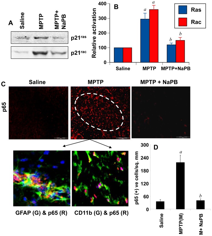
Figure 6. Activation of small G proteins (p21ras and p21rac) and NF-κB in ventral midbrain of MPTP-intoxicated mice is NaPB-sensitive.
A) Mice were treated with NaPB (200 mg/kg body wt/d) via gavage from 1 d prior to MPTP injection. Six h after the last injection of MPTP, activation of p21ras and p21rac was monitored in ventral midbrain tissues. Experiment was repeated three times each time using two animal in each group. B) Bands from three different mice were quantified and activation of p21ras and p21rac is shown as percent of control. C) Mice were treated with NaPB (200 mg/kg body wt/d) from 3 h after the last injection of MPTP. Twenty-four h after the last injection of MPTP, ventral midbrain sections were immunostained for p65 (low magnification). Midbrain sections of MPTP-intoxicated mice were also double-labeled for p65 and glial cell markers (GFAP for astrocytes and CD11b for microglia). Results represent three independent experiments. D) NF-κB p65 positive cells counted in four nigral sections (two images per slide) from each of four mice in an Olympus IX81 fluorescence microscope using the MicroSuite™ imaging software are mentioned as cells/mm2. ap<0.0001 vs saline-control; bp<0.0001 vs MPTP.

