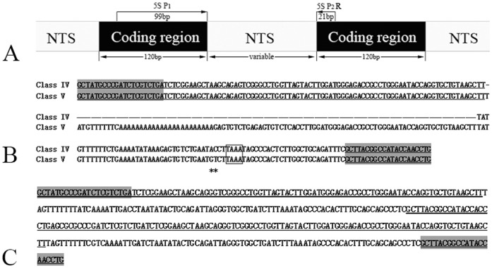
Figure 5. Representative sequences of monomeric 5S rDNA and dimeric 5S rDNA.
(A) Arrangement of eukaryotic 5S rDNA and the illustration of the PCR amplification with 5S primers P1 and P2R; (B) Representative sequences of 5S rDNA Class IV and Class V from TC; (C) The dimeric 5S rDNA tandem arrays (NTS–I–N) of 4nRT hybrids. The gene sequences of 5S rDNA are underlined and the shaded regions show the 5S primers. Dashes indicate alignment gaps; asterisks represent variable sites; TATA element (TAAA) is included in box.

