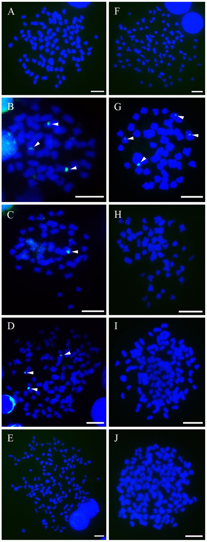
Figure 8. Fluorescence photomicrographs of mitotic metaphase chromosomes of RCC, TC and their hybrid offspring.

Signals were detected with fluorescein isothiocyanate (FITC)-conjugated avidin and all the metaphase chromosomes were stained with DAPI. A–E (mitotic metaphase chromosomes of RCC, TC, 2nRT, 3nRT and 4nRT hybrids respectively) show the single-label FISH results hybridized with a probe from the cloned repeated fragments of class IV; F–J (mitotic metaphase chromosomes of RCC, TC, 2nRT, 3nRT and 4nRT hybrids respectively) demonstrate the hybridization results after single-label FISH with a probe from the cloned repeated fragments of class V. The white arrowheads indicate the fluorescent signals (green) of 5S rDNA. Scale bar in A–J, 3 µm.
