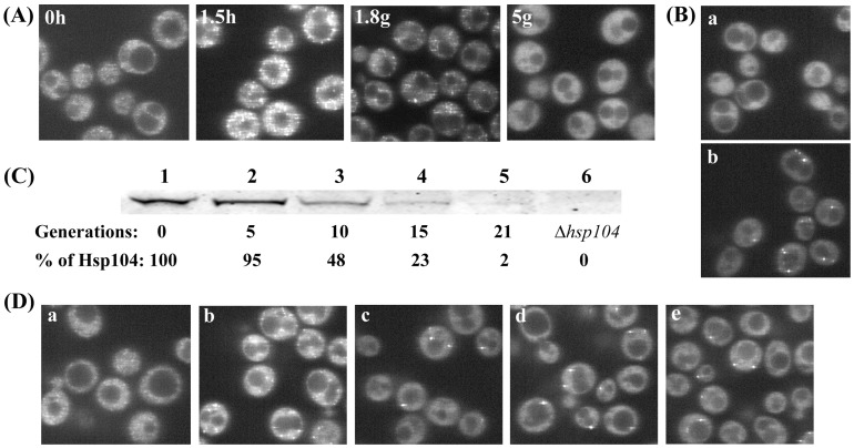
Figure 2. Effect of guanidine on fluorescence of NGMC foci in [PSI+] yeast in presence and absence of Hsp104.
(A) Change in fluorescence of NGMC during curing by guanidine. Cells were grown in SD with 5 mM guanidine for the indicated growth time in hours (h) or generations (g). (B) Fluorescent imaging of [PSI +] cells either with the empty vector (a) or plasmid pFL39-GAL-HSP104KT expressing Hsp104-2KT (b) were grown for 5 generations in SGal with 5 mM guanidine. (C) Western blot analysis of Hsp104 to determine the level of Hsp104 following excision of Hsp104 after inducing expression of Flp recombinase. Yeast were grown in SGal to induce expression of Flp recombinase to delete the FRT-sites flanked HSP104. Lysates were obtained from the conditional deletion yeast strain of HSP104 grown in SD (lane 1) and SGal for indicated generations (lane2–5), and the Δhsp104 strain (lane 6). The percent Hsp104 calculated from the intensity at each time relative to the intensity of the control was corrected for protein loading using the Pgk1 standard. (D) Fluorescent microscopic images of yeast expressing NGMC before and after conditional deletion of HSP104. The conditional deletion yeast strain of HSP104 was grown in SD (a) or in SGal for 10 generations (b), followed by another 5 generations in SGal with (c) and without (d) 5 mM guanidine. Cells in panel (d) were incubated for 1 h in water (e). Images show one slice from a Z-stack (16 slides, interval 0.4 µm).

