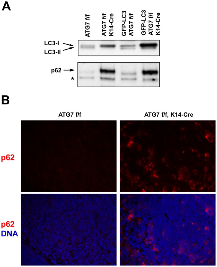
Figure 2. Deletion of ATG7 in K14-expressing cells suppresses autophagy and leads to accumulation of p62 in the thymus.
(A) Thymus lysates of the ATG7 f/f and ATG7 f/f K14-Cre, either carrying the GFP-LC3 transgene or not, were subjected to Western blot analysis for LC3 (upper panel) and p62 (lower panel). Bands corresponding to LC3-I and LC3-II (indicative of active autophagy) are marked by arrows. Note the presence of LC3-II bands in ATG7 f/f thymi and the accumulation of LC3-I as well as p62 in ATG7 f/f K14-Cre mice. The position of an unspecific band, that shows equal loading of the lanes, is marked by an asterisk. (B) Immunofluorescence analysis of p62 expression (red) in the thymus. Note the accumulation of p62 in the characteristically shaped epithelial cells of ATG7 f/f K14-Cre mice. Nuclei are labeled with Hoechst 33258 (blue). The complete field of view under 200-fold magnification is shown in each panel.

