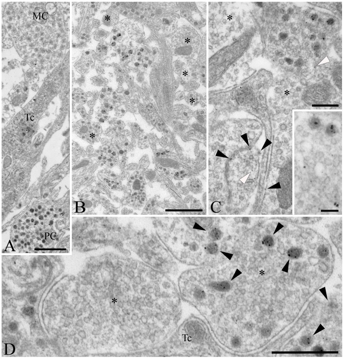
Figure 2. Ultrastructure of D3 immunoreactive elements in the rat mediobasal hypothalamus.
(A) D3 immunoreactivity, identified by silver grain deposits, appear primarily in axon varicosities containing dense core vesicles of 80–120 nm diameter in the upper external zone of the median eminence, characteristic of axons of parvocellular (PC) neurons. No or a few silver grains could be observed in association with organelles of magnocellular neurons (MC) or tanycytes (Tc), respectively. (B) D3-positive axons exhibiting various degrees of labeling are mixed with non-labeled fibers (asterisks) in the external zone of the median eminence. (C) Although silver grains occasionally appear in association with the plasma membrane (black arrowheads) and with small clear vesicles (white arrowheads) of the axon varicosities, the majority are not labeled (asterisks). (D) In contrast, the dense core vesicles accumulate most the reaction product (black arrowheads), as visible at high power magnification in the vicinity of the capillaries of the external zone of the median eminence. Unlabeled small clear vesicles are indicated with asterisk. Tc, tanycyte; Scale bars: 1 µm in A–B, 250 nm in C, 100 nm in inset on C, 500 nm in D.

