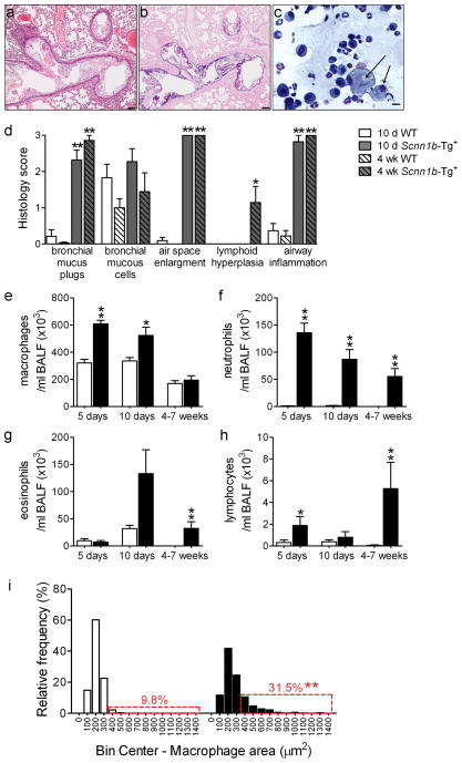
Figure 7. Germ-free (GF) Scnn1b-Tg+ mice develop lung inflammation similar to Scnn1b-Tg+ mice raised in conventional SPF conditions.
(a, b) Representative photomicrographs of left lobe main stem bronchus from 6 week-old GF Scnn1b-Tg+ mice, illustrating alveolar space enlargement, mucus obstruction, and airway inflammation. H&E (a) and AB-PAS (b) stain. Scale bar 100 μm. (c) Representative photomicrograph of BAL cytospin preparation from GF Scnn1b-Tg+ mice, illustrating mucus plugs (light blue), granulocytes and large/foamy macrophages (arrows). Giemsa stain, scale bar = 20 μm. (d) Semi-quantitative histopathology scores for 10 day old (10 d, open bars) and 4 week-old (4 wk, hatched bars) Scnn1b-Tg+ mice (gray) and WT littermates (white) raised in GF conditions, n= 11 Scnn1b-Tg+ and 11 WT littermates at 10 days, n=7 Scnn1b-Tg+ and 9 WT littermates at 4 weeks of age. T test ** p<0.005, * p<0.05 vs. WT littermates. (e-h) Longitudinal differential BAL cell counts for GF Scnn1b-Tg+ mice (■) and WT littermates (□). n= 11 and 11 at 5 days, n= 18 and 8 at 10 days, n= 8 and 7 at 4–7 weeks, for GF WT and GF Scnn1b-Tg+ mice, respectively. (i) Macrophage size distribution in 4–7 week-old GF Scnn1b-Tg+ mice (■, n=7) and WT littermates (□, n=8). Boxed regions highlight the percentage of total macrophages larger than the 90th percentile in WT mice. T test ** p<0.005 vs. WT littermates.

