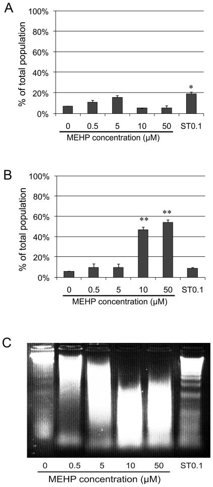
Figure 5. Effects of MEHP on cell death.
Apoptosis/necrosis assay (Invitrogen/Molecular Probes) was performed after exposure to increasing doses of MEHP (0, 0.5, 5, 10, 50 μM) for 10 h or to 0.1 μM Staurospaurine (positive control ST0.1). Following treatment, cells positively stained by Yo-Pro and/or propidium iodide (PI) were counted using Tali™ image based cytometer. Cells positive for Yo-Pro only were apoptotic (A), while PI positive cells were considered necrotic (B). TALI™ counts were standardized over the vehicle control. Data are expressed as the means of 3 experiments in duplicates, and error bars represent the standard error to the mean. * indicates a p-value <0.01, ** indicates a p-value <0.005. C: apoptosis was evaluated by assessing the presence of DNA laddering by gel electrophoresis. Results indicated that MEHP did not significantly trigger an increase of apoptosis, but significantly increased necrosis for the highest doses (10 and 50 μM). Staurosporine (0.1μM) triggered significant apoptosis, which is shown by DNA laddering.

