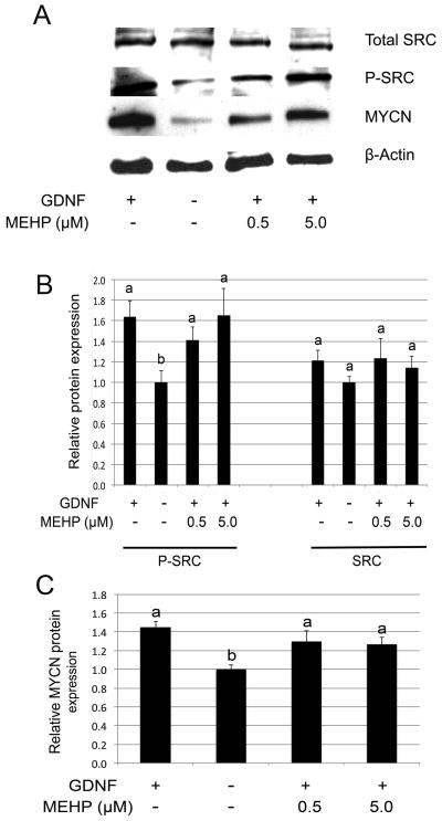
Figure 6. Influence of MEHP on GDNF-SRC signalling.
Western Blot analysis of SRC protein phosphorylation and MYCN protein expression was performed after 10 h exposure to MEHP (0, 0.5 and 5μM), followed by stimulation with GDNF for 20 min (phospho-SRC analysis) or 18 h (MYCN analysis). Beta-actin was used as loading control, and band intensities where standardized over the vehicle control as reference. Figure A represents a typical Western blot, and Figures B and C represent quantification of the band intensities. Neither GDNF-dependent SRC phosphorylation (A and B), nor GDNF-dependent MYCN protein expression (A and C) were significantly impaired by the presence of MEHP.

