Figure 2. Inhibition of hydrogen peroxide-mediated Akt signaling by 4-HNE.
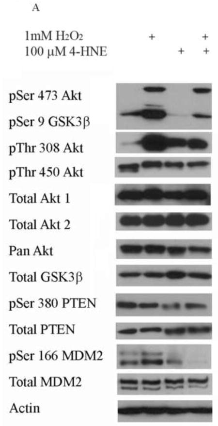
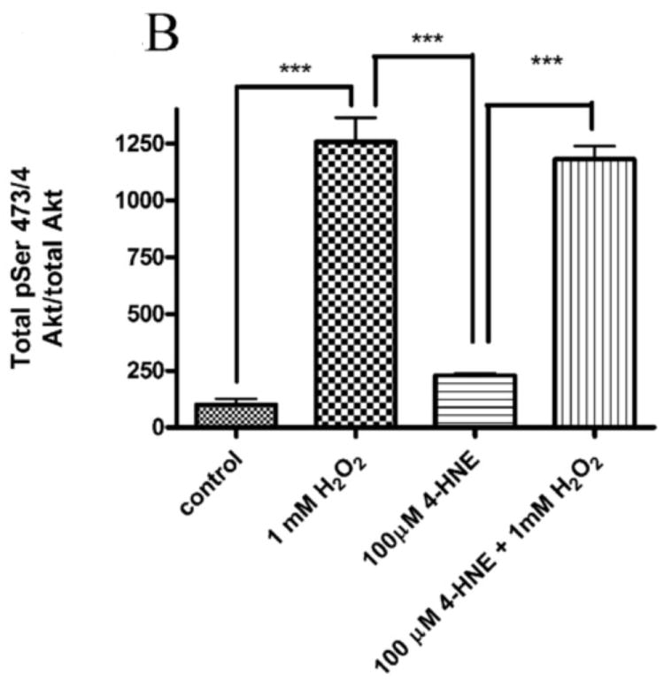
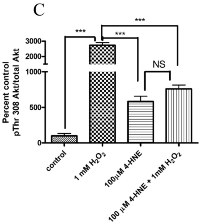
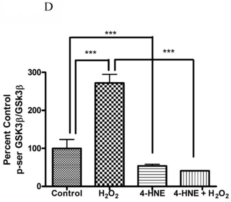
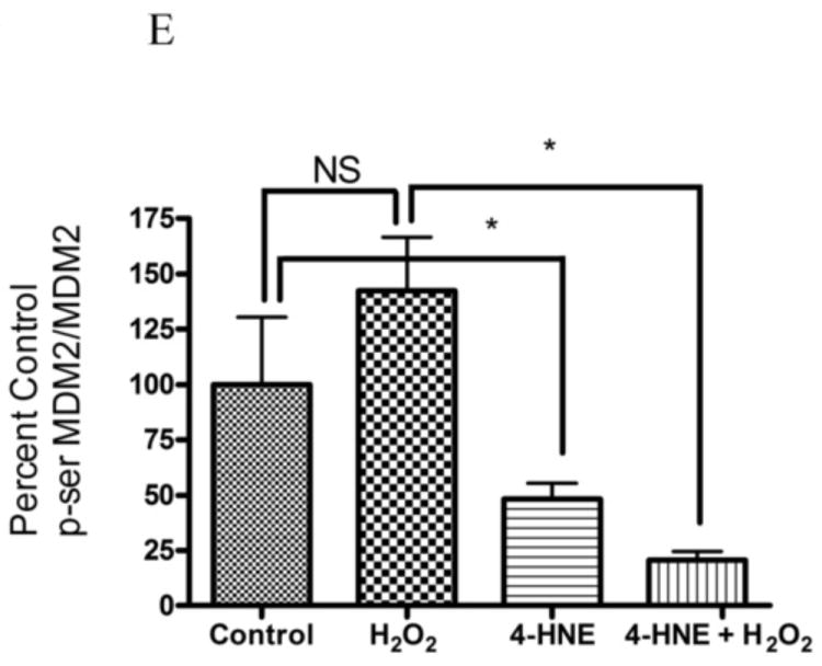
Cells were treated in serum free media with 100μM 4-HNE or control for 60 min, washed in serum free media and stimulated with 1mM hydrogen peroxide for 5 minutes. Cells were lysed and processed as stated in methods. (A) Western blot using antibodies for the following proteins: Akt (Ser473/4), Akt (Thr 308/9), Akt (Thr 450), Akt1, Akt2, PTEN, PTEN (pSer380), p-GSK3β (Ser9), GSK3β, p-MDM2 (Ser166), MDM2 and actin (Note: Film exposures for pSer473 Akt are less than 2 seconds). Each blot is representative of 3 independent experiments, for MDM2 samples were run on a 5% SDS PAGE gel, all other samples were run on a 7% SDS PAGE. (B) Quantification of pSer473/4 Akt. (C) Quantification of pThr 308/9 Akt. (D) Quantification of pSer 9 GSK3b. (E) Quantification of pSer 166 MDM2. Statistical analysis was via 1-way analysis of variance with Tukey’s multiple comparison test *p<0.05, ***p<0.001.
