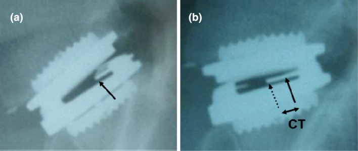
Fig. 3.
Radiographs a in flexion and b in extension of a mobile-core prosthesis. Arrows localize the anterior part of the marker inserted in the core: the full one in flexion and the dotted one in extension. The difference of location between the two radiographs corresponds to the core translation (CT)

