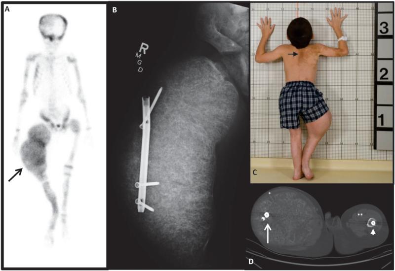
Figure 1. Initial Clinical Presentation.
A) 99Technectium-MDP bone scan showing extensive tracer uptake at multiple areas of FD and ballooning expansion of the right femur (arrow). B) X-ray shows massive expansion of right femur and displacement of a rod that was originally in the intramedullary canal. C) Photograph showing classic “coast-of-Maine” café-au-lait spots (arrow) and the effect of massive overgrowth of the right femur. D) CT at the level of the femura mid-shafts, with lateral displacement of the rod on the right (white arrow), and an intramedullary rod on the left (arrow head). The quadriceps muscles on the right have been stretched to dysfunctional bands (*), compared to the left (**).

