
Figure 3. Runx1 controls the stromal and cartilaginous callus formation at the early stage of bone fracture healing.
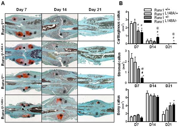
Seven to 9 week old mice were used for the femoral fracture experiments as described in the materials and methods section. (A) For histological analysis, the fracture calluses were stained with Safonin O (Red: cartilage) and Fast Green (Green: mineralized tissue) to identify the stromal (•), cartilaginous (*), and mineralized (#) callus formation. (B) Histomorphometric assessment was also performed to measure the size of the stromal, cartilaginous, and bony callus at day 7, 14, and 21 post fracture. n=4-7 mice per group. *p<0.05 vs Runx1 +/+; #p<0.05 vs Runx1 L148A/+; and @p<0.05 vs Runx1 +/−.