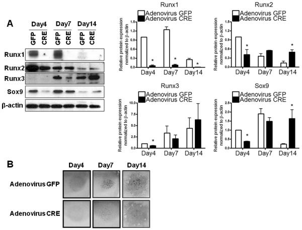
Figure 6. Runx1 regulates the chondrogenic differentiation of the early mesenchymal progenitor cells in vitro.

Pure mesenchymal stem cell populations isolated from E12.5 limb buds of Runx1F/F were infected with adeno-Cre virus and then cultured as micromass. Adeno-GFP virus was used as a control. (A) Nuclear fractions isolated from micromass culture were used for protein analyses of Runx1, Runx2, Runx3, Sox9, and β-actin at day 4, 7, and 14. Three independent experiments were performed to measure protein levels of Runx1, Runx2, Runx3, and Sox9. Their band intensities were quantified by NIH ImageJ. Values are expressed as means ± SE, normalized to β-actin. *p<0.05 vs adeno-GFP infected controls at the same time point. (B) Micromass cultures were stained with Alcian blue at the time points indicated above.
