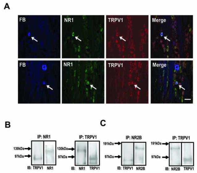
Figure 2.
NR1 and TRPV1 expression and co-immunoprecipitation in TG. (A) immunohistochemical staining of TG sections showing FB-labeled muscle afferents, TRPV1- and NMDA-labeled neurons as indicated. The arrows indicate muscle afferents that co-express both NR1 and TRPV1. Scale bar: 50 μm. (B) (left) Immunoblot (IB) using anti-TRPV1 or anti-NR1 antibody following immunoprecipitation (IP) of TG extract with anti-NR1 antibody. (Right) Reverse IP with anti-TRPV1 antibody and IB with anti-NR1 or anti-TRPV1 antibody. (C) The same IB-IP protocols were used to show co-immunoprecipitation of TRPV1 and NR2B subunit.

