
Figure 5.
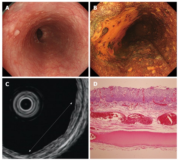
Findings for an m1 cancer of the esophagus. A: Endoscopic features. A reddish depressed lesion was located on the anterior and left wall of the middle esophagus; B: Endoscopic features after iodine dye. Biopsy specimens showed squamous cell carcinoma; C: Endoscopic ultrasound (EUS) features. The white dotted line indicates the extent of the lesion. EUS revealed an irregularity of the first layer and a slight thickness of the second layer; D: Pathological findings. The tumor was confined to the epithelium. (Hematoxylin and eosin stain, × 40).