
Figure 9.
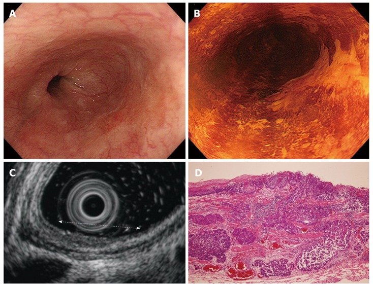
Findings for an sm2 cancer of the esophagus. A: Endoscopic features. A depressed lesion was located on the posterior and right wall of the middle esophagus; B: Endoscopic features after iodine dye. Biopsy specimens showed squamous cell carcinoma; C: Endoscopic ultrasound (EUS) features. The white dotted line indicates the extent of the lesion. EUS revealed a thickness of the second layer and a disappearance of the third and fourth layer. The fifth layer had become thin, but the sixth layer was intact; D: Pathological findings. The tumor was invading the submucosal layer to a 320 μm depth. (Hematoxylin and eosin stain, × 40).