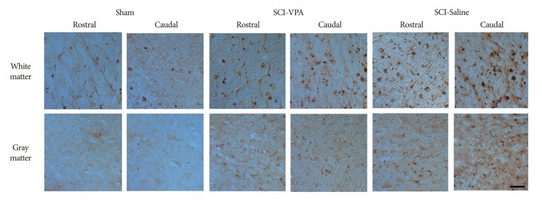
Fig. 6.
Representative photographs of ED-1 immunoreactive cells from SCI to sham animals at 6 mm both rostral and caudal to the lesion epicenter, 40×. Considerable decline of the immunoreactivity of ED-1 is evident in both white and gray matter in VPA-injected groups, while in saline-injected groups high immunoreactivity of ED-1 is evident. Scale bar=50 µm; 40× magnification. SCI : spinal cord injury, VPA : valproic acid.

