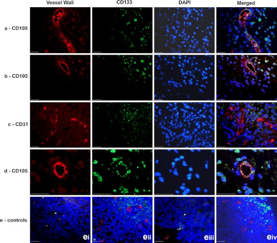
Fig. 3.

Co-immunofluorescence staining (blue nuclear DAPI staining): a CD105 (red) with CD133 (green) in an anaplastic astrocytoma, b CD105 (red) with CD133 (green) in a glioblastoma multiforme, c CD31 (red) with CD133 (green) in a glioblastoma multiforme, d CD105 and CD133 demonstrating co-staining of individual cells in vessel wall (arrowed) in a glioblastoma (scale bars all 25 μm). e Control sections of human tonsil demonstrating negative (ei) and positive (eii) controls for CD105 (red) and CD133 (green) and negative (eiii) and positive (eiv) controls for CD31 (red) and CD133 (green)
