
Fig. 4.
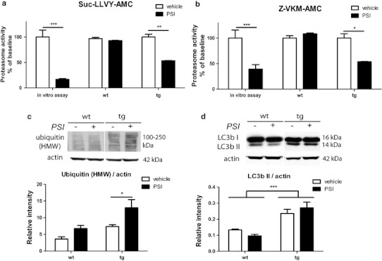
Effects of systemic PSI treatment on the ubiquitin–proteasome system and the autophagy–lysosomal pathway in the brain. a Proteasome activity given as a percentage of baseline fluorescence of substrate III (Suc-LLVY-AMC) was reduced by about 93 % after in vitro application of PSI (in vitro assay) and reduced by about 50 % after in vivo application of PSI in transgenic (tg) mice at 2 weeks after first intoxication. However, no effect of PSI treatment on brain proteasome activity was measured at the same time point in wild-type (wt) mice. b Proteasome activity given as a percentage of baseline fluorescence of substrate IV (Z-VKM-AMC) was reduced by about 61 % after in vitro application of PSI (in vitro assay) and reduced by about 50 % after in vivo application of PSI in tg mice at 2 weeks after first intoxication. No effect of in vivo PSI treatment on brain proteasome activity was measured in wt mice at the same time point. c Representative immunoblot showing levels of polyubiquitinated high molecular weight (HMW) species as well as densitometric analysis of HMW ubiquitin smear (100–250 kDa) in all mouse groups presented as relative intensity to actin. d Representative immunoblot showing levels of LC3B I (16 kDa) and LC3B II (14 kDa) as well as densitometric analysis of LC3B II in all mouse groups presented as relative intensity to actin. Data are presented as mean ± SEM. In all cases n = 3. Data were analyzed by two-way ANOVA with post-hoc Bonferroni’s test. *p < 0.05; **p < 0.01; ***p < 0.001