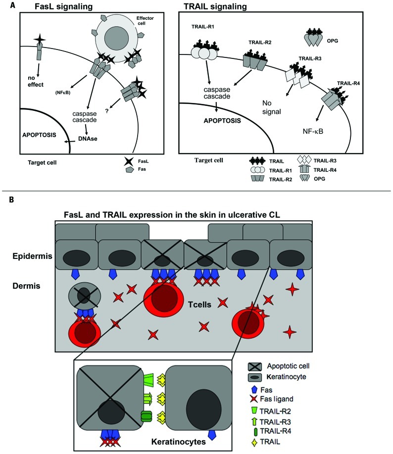
FIGURE 2.
FasL and TRAIL signaling in ulcerative cutaneous leishmaniasis. (A) FasL and TRAIL signaling induce apoptosis or proinflammatory signaling depending on the receptor expression on the target cell. (Left) Fas is expressed as a homodimer on cell-surfaces. Upon FasL ligation to trimerized Fas caspase activation leading to apoptosis is initiated. Fas may also lead to NF-κb activation. sFas probably serves as a negative modulator of FasL. The function of sFasL is debated and it has been suggested that the concentration will determine if apoptosis can be triggered through Fas by sFasL. (Right) Apoptosis is induced through TRAIL activation of TRAIL-R1 and -R2. TRAIL-R3 is a decoy receptor and will not initiate intracellular signaling. Activation of TRAIL-R4 leads to NK-κB activation. OPG is a soluble receptor binding TRAIL and its function has not been elucidated. (B) A model for keratinocyte apoptosis in the ulcerative process of CL. Keratinocytes upregulate Fas, TRAIL-R2, -R4, and TRAIL as a result of the inflammatory reaction surrounding L. major-infected macrophages. FasL-expressing T cells and TRAIL-expressing inflammatory cells accumulate at the site of L. major infection. The Fas–FasL/Fas–sFasL and TRAIL/TRAIL-R interaction may lead to keratinocyte apoptosis and ulceration as well as to activation induced T cell apoptosis.

