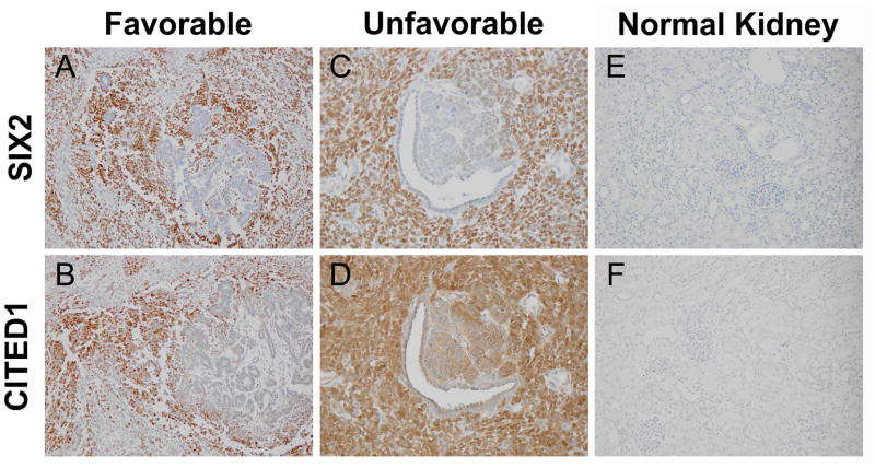
Figure 1.
Serial sections from a favorable histology WT show blastemal immunopositivity for SIX2 (A) and CITED1 (B). Epithelia and stroma are immunonegative, similar to the embryonic kidney staining pattern. Magnification = 20X. Serial sections from unfavorable histology WT with similar overlap in blastemal expression of SIX2 (C) and CITED1 (D). Areas surrounding epithelial differentiation are weakly CITED1 positive, but SIX2 negative, demonstrating divergent staining patterns unique to a proportion of tumors examined. Magnification = 40X. Normal kidney controls show immunonegativity for SIX2 (E) and CITED1 (F). Magnification = 20X.

