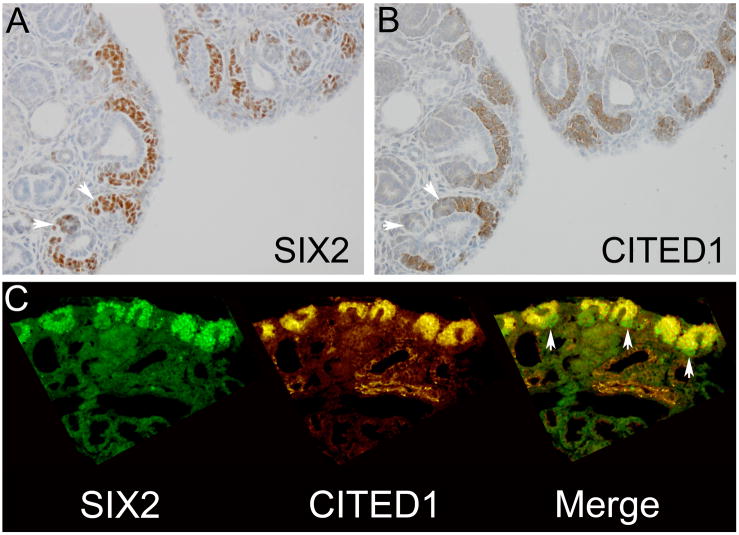
Figure 2.
Serial sections from e18.5 mouse fetal kidney show a population of cells in the ventral cap mesenchyme and in pre-tubular aggregates (arrowheads) that are SIX2 positive (A), but CITED1 negative (B). Magnification = 20X. Double immunofluorescence for SIX2 and CITED1 on the same section (C) identifies this SIX2 positive, CITED1 negative population in the ventral cap mesenchyme (arrowheads). Magnification = 20X.

