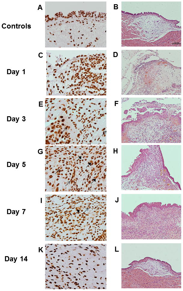
Figure 1. High-mobility group chromosomal box (HMGB1) expression following vocal fold surgical injury.

(A, C, E, F, I and K) Representative H&E staining of uninjured and injured vocal fold morphology over 2 week post surgery (200X magnification). (B, D, F, H, J and L) Representative IHC staining of HMGB1 of the same rats (600X magnification) in corresponding H&E micrographs. HMGB1 is stained brown. Positive nuclear staining was evident in uninjured vocal folds. In addition to the nuclear expression, cytoplasmic (thin arrows) and extracellular (thick arrows) HMGB 1 staining was evident in injured samples from Day 1 to Day 7 post surgery. In Day 14, cytoplasmic and extracellular HMGB1 deposition became less evident.
