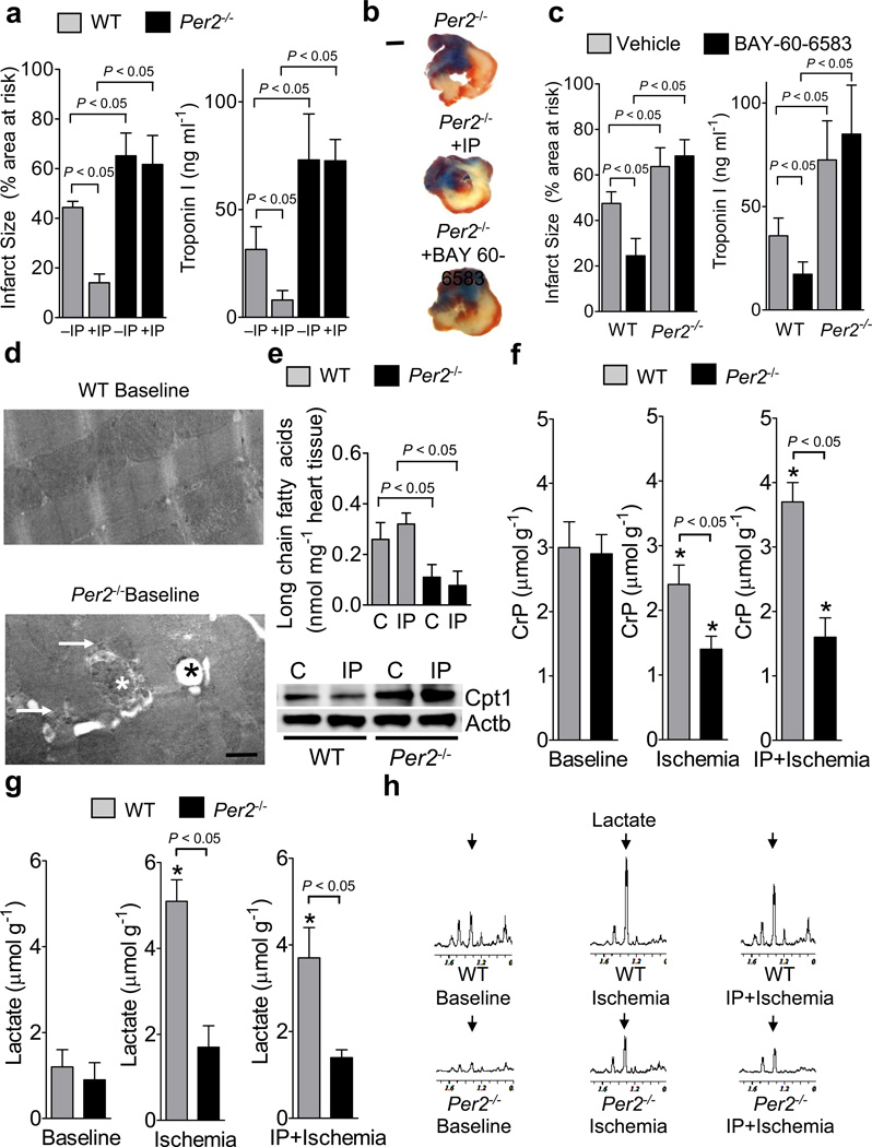
Figure 3. Functional role of Period 2 during myocardial ischemia and ischemic preconditioning.
(a–c) Per2−/− mice or littermate controls matched in age, weight and gender were exposed to 60 min of in situ myocardial ischemia followed by 2h or reperfusion, or received IP pretreatment or Adora2b agonist (BAY 60-6583) treatment prior to myocardial ischemia. IP consisted of 4 cycles of 5 minutes of myocardial ischemia and 5 minutes of reperfusion. Infarct sizes are expressed as the percent of the area at risk that was exposed to myocardial ischemia. In parallel, measurements of the myocardial injury marker troponin I were performed (mean±SD; n=6). (b) Representative infarct staining from Per2−/− mice exposed to 60 min of ischemia, and 2h reperfusion alone (−IP), or additional IP or Adora2b agonist treatment (+IP/BAY 60-6583,c) are displayed (blue indicates retrograde Evan’s blue staining; red and white: area at risk; white: infarcted tissue; scale bar represents 50 µm). (d) Electron microscopy from wildtype and Per2−/− heart tissue. Baseline wildtype and Per2−/− mice showed normal sarcolemal structures, however, in some areas Per2−/− mice exhibited enhanced glycogen content (white arrow), swollen mitochondria (white star) and lipid accumulation within mitochondria (black star); scale bar represents 500 nm. (e) Per2−/− mice or littermate controls matched in age, weight and gender were subjected to in situ IP treatment consisting of 4 cycles of IP (5 minutes of ischemia, 5 minutes of reperfusion). Cardiac preconditioned tissue was shock-frozen and analyzed for long chain fatty acids using an enzymatic ELISA KIT from Biovision (mean±SD; n=3) and protein levels of carnitine-palmitoyltransferase 1 (Cpt1). One representative blot of three is displayed. (f–h) Per2−/− mice or littermate controls matched in age, weight and gender were exposed to 60 min of in situ myocardial ischemia with or without ischemic preconditioning (IP; 4 cycles of 5 min ischemia followed by 5 min of reperfusion) prior to myocardial ischemia. For nuclear magnetic resonance (NMR) analysis of cardiac metabolites, cardiac tissue was shock frozen immediately after ischemia. (f) Creatinephosphate (CrP) levels in wildtype and Per2−/− mice using NMR. Note: IP mediated conservation of CrP storage is abolished in Per2−/− mice. (g, h) Lactate levels and corresponding NMR spectra. (n=3, * significant changes compared to baseline conditions, p<0.05).

