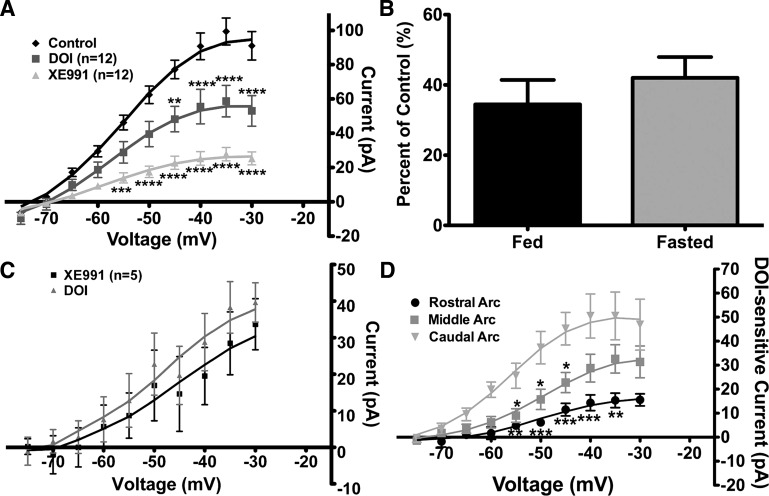
Fig. 5.
DOI suppresses the M-current in fasted male mice similar to fed male mice, and expression of DOI-sensitive M-current is regionally dependent in arcuate POMC neurons. A: in fasted male mice, 20 μM DOI suppressed M-current activity. After DOI treatment, 20 μM XE-991 was perfused for 10 min. Current-voltage plots were analyzed by 2-way ANOVA (P < 0.0001, F = 17.5, df = 2) followed by Bonferroni-Dunn multiple comparison tests: **P < 0.01; ***P < 0.001; ****P < 0.0001 vs. control. B: percentage of control current suppressed by DOI at −35 mV in POMC neurons from fed and fasted male mice. C: DOI did not significantly suppress more M-current after XE-991 perfusion in POMC neurons from fed male mice. D: amount of M-current suppressed by DOI (DOI-sensitive current) is dependent on the region of the arcuate (Arc) nucleus. Suppression of the M-current was more robust in the caudal (n = 7 cells) and middle (n = 16 cells) regions than in the rostral region (n = 8 cells). Current-voltage plots were analyzed by 2-way ANOVA comparing arcuate regions (P < 0.01, F = 7.3, df = 2) followed by Bonferroni-Dunn multiple comparison tests: *P < 0.05; **P < 0.01; ***P < 0.001 vs. caudal.

