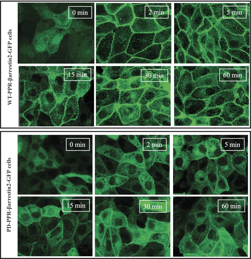
Fig. 2.
Decreased PTH-induced β-arrestin2-GFP membrane recruitment in the PD-PPR cells stably expressing β-arrestin2-GFP compared with the WT-PPR cells. The WT-PPR-βarrestin2-GFP and PD-PPR-βarrestin2-GFP cells were grown on cover slips in 6-well plates for 48 h. The cells were then treated with 100 nM PTH (0–60 min at 37°), washed with ice-cold PBS, and fixed with 4% paraformaldehyde for 20 min at RT, mounted, and examined under a confocal microscope. Images shown were taken at the X-Y horizontal planes (Z-series) in the middle of the cells. This experiment was repeated 3 times with similar results. These experiments were performed in the GFP-untagged WT-PPR and PD-PPR cells.

