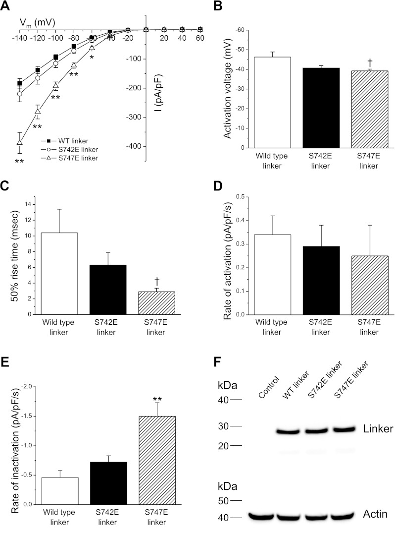
Fig. 6.
Effect of expression of wild-type, S742E, or S747E linkers on functional properties of wild-type CLH-3b. Cells were cotransfected with 1.5 μg, 0.075 μg, and 4 μg of wild-type CLH-3b, GCK-3, and linker cDNAs, respectively. A: whole cell current amplitude and current-to-voltage relationships. B: activation voltages. C: 50% rise time. D and E: rates of swelling-induced activation (D) and shrinkage-induced inactivation (E). Values are means ± SE (n = 10–13). †P < 0.05, *P < 0.01, **P < 0.001 compared with cells expressing wild-type linker. F: Western blot showing expression levels of V5-tagged wild-type, S742E, and S747E linkers. Lanes were also probed with an anti-actin polyclonal antibody to assess protein loading. Control, nontransfected cells.

