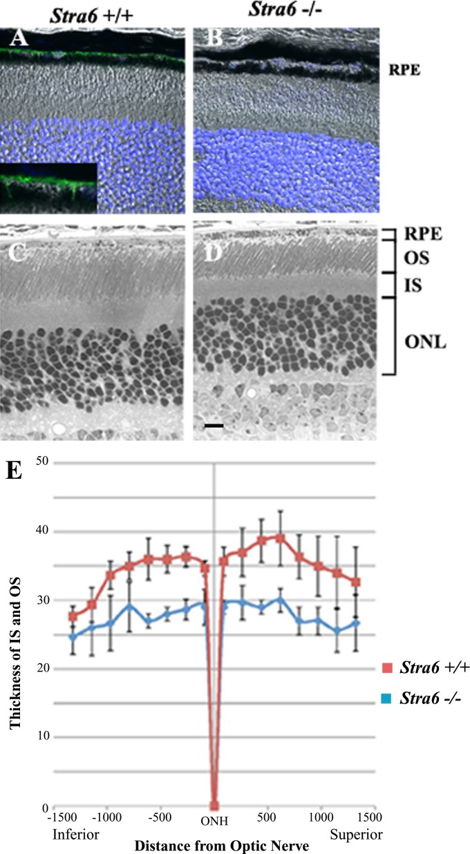
Figure 3.
Histological features of the retina in stra6 mutants and controls. (A, B) Frozen sections (10 μm) of retina from stra6 +/+ and stra6 −/− mice. STRA6 staining was performed by using the anti-STRA6 antibody from abCam Inc. at 1:200 dilution. (C, D) Images of 1 μm plastic sections of retina from both genotypes showing the shortening of outer segments and inner segments in the retina of mice lacking STRA6 (D). (E) Measurements of outer/inner segment thickness along the entire extent of the retina. The optic nerve head (ONH) was used as a point of reference to differentiate the superior (right) and inferior (left) hemispheres. Means of four representative retinas are shown for each stra6 +/+ and stra6 −/− genotype. Eight different points were measured in each hemisphere. ONL, outer nuclear layer. Bar, 20 μm.

