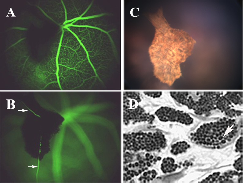
Figure 4.
Representative fundus analysis and light microscopy images from a 6-week-old stra6 −/− mouse are shown. (A, B) The presence of a PHPV structure in two different focal planes observed following IP injection of fluorescein. Vascularization of these structures is observed by the presence of blood vessels (B, arrows). (C) The asteroid-like appearance of the PHPV structure from (A) and (B) in standard fundus imaging. (D) Epon-Araldite sagittal section (1 μm) showing the internal features of the PHPV body, which includes pigmented melanocytes.

