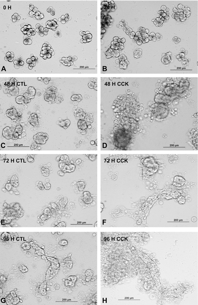
Fig. 1.
Morphology of isolated pancreatic acinar cultures. Mouse isolated acini were plated in 24-well plates coated with collagen and imaged at 0 h (A), 24 h (B), 48 h (C and D), 72 h (E and F), or 96 h (G and H). CCK (1 nM) was added at 24 h to wells shown in D, F, and H. Live cells were imaged through the plastic culture well with an inverted microscope via a ×20 objective and recorded digitally. The acini are attached at 24 h and have flattened and begun to spread on the surface at 48 h. Spreading thereafter is enhanced by CCK. CTL, control.

