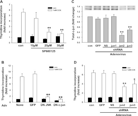
Fig. 8.
Inhibition of CCK-induced pancreatic acinar cell proliferation by JNK inhibitor, dominant-negative JNK, dominant-negative c-Jun, and c-Jun short hairpin RNA (shRNA). A: effect of JNK inhibitor SP600125 on acinar cell [3H]thymidine incorporation. Acinar cells were preincubated with the inhibitor at the indicated concentration for 1 h followed by addition of 1 nM CCK. Values are means ± SE of 3–5 experiments. **P < 0.01 compared with control CCK-treated groups. B: effect of dominant-negative (DN) JNK and dominant-negative c-Jun on acinar cell [3H]thymidine incorporation. Adenovirus (Adv; 107 pfu/ml) was added when acinar cells were plated; 1 nM CCK was added 24 h later. Values are means ± SE of 3–5 experiments. C and D: reduction of CCK-induced acinar cell proliferation by c-Jun shRNA. Adenovirus was added at 107 pfu/ml when acinar cells were plated. C: cells were cultured for 24 h followed by protein extraction. The efficiency of shRNA to knockdown c-Jun expression was evaluated by Western blot. NS, nonspecific. **P < 0.01 compared with control group without any adenovirus infection. D: effect of c-Jun shRNA on CCK-induced acinar cell [3H]thymidine incorporation. For C, duplicate culture wells were run on gels and imaged, and after quantification the image was spliced to show only 1 lane per condition as described in materials and methods. For this reason a white line is placed between the lanes. **P < 0.01 compared with control (no virus) CCK-treated groups. Values are means ± SE of 3–5 experiments. **P < 0.01; *P < 0.05 compared with CCK-treated group without any adenovirus infection.

