Fig. 3.
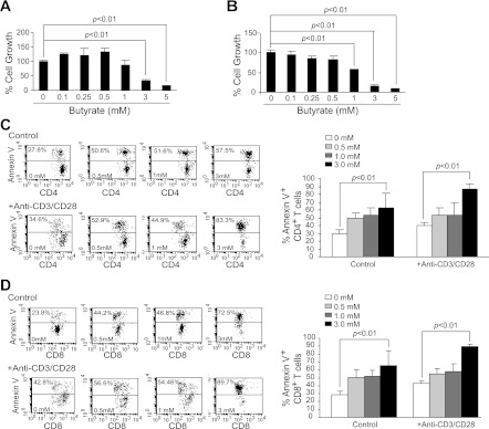
Butyrate induces T cell apoptosis. Purified CD4+ (A) and CD8+ (B) T cells were cultured in anti-CD3/CD28 mAb-coated plates for 24 h. Butyrate was then added to the culture for another 24 h. Cellular proliferation/survival was analyzed using MTT assays. Cellular proliferation in the absence of butyrate was set as 100%. C: spleen cells were cultured in the absence (control) and presence (+Anti-CD3/CD28) of anti-CD3/CD28 mAbs for 24 h. Various concentrations of butyrate were then added to the cultures for another 24 h. Cells were collected and stained with Annexin V and CD4 mAb and analyzed by flow cytometry for apoptosis. Left: apoptosis profiles of CD4+ T cells are shown. Right: apoptotic cell death of CD4+ T cells was quantified by the formula: % Annexin V cells = % Annexin V-positive cells in the presence of butyrate − % Annexin V-positive cells in the absence of butyrate. Shown are representative results from 1 of 3 experiments. D: CD8+ T cells apoptosis in response to butyrate was analyzed as in C.

