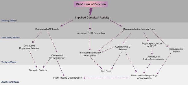
Figure 7. Schematic representation of upstream and downstream defects caused by PINK1 deficiency.
As discussed in the text and illustrated in this figure, Complex I deficiency can explain almost any alteration previously found to be associated with PINK1 loss of function. The decreased electron transfer at the level of Complex I can lead to reduced Δψm (present study), decreased ATP generation (Clark et al, 2006) and increased ROS production (Gautier et al, 2008). Deficient ATP levels affect dopamine release (Kitada et al, 2007) and RP mobilization (present study) causing reduced RP mobilization. Additionally, reduced Δψm can destabilize cytochrome c release (present study), and dephosphorylation of Drp1 that causes alterations in mitochondrial fusion/fission rates and morphology (Cereghetti et al, 2008). Recruitment of Parkin to such mitochondria (Narendra et al, 2008) will partially act protectively, as it will remove the most severely affected mitochondria, also explaining the genetic interaction between Parkin and Pink (Poole et al, 2008).

