
Figure 2. Histological features of EMT in cancer and fibrosis.
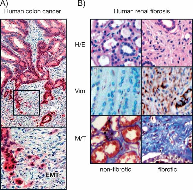
- Immunohistochemical staining of a colorectal carcinoma for β-catenin in red and nuclear counterstaining in blue. The central area of the tumour exhibits polarized epithelial tumour cells lacking nuclear β-catenin, while cells at the invasive front undergo EMT and show nuclear β-catenin (Brabletz et al, 2001). Picture from Dr Thomas Brabletz, Univ. Freiburg (Germany).
- Histological sections showing the halmarks of EMT in the medulla of fibrotic kidney from patients subjected to nephrectomy. Sections are stained with haematoxylin–eosin (H/E) to better appreciate cell morphology and the disappearance of the tubular structures in fibrosis, vimentin expression showing mesenchymal cells (brown, Vim) and fibrotic deposits revealed by the blue Masson–Trichome staining (M/T) (Boutet et al, 2006).