Fig. 5.
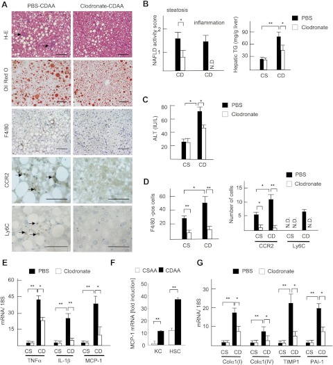
Kupffer cell depletion delays the progression of steatohepatitis. Mice were treated with liposomal PBS (closed bar) or clodronate (open bar), and fed on the CSAA (CS) and CDAA (CD) diets for 2 wk. A: H-E, Oil red O staining, and immunohistochemical staining for F4/80, CCR2, and Ly6C. Steatosis and inflammatory cell infiltration (arrows) are attenuated by clodronate liposome injection. Bar = 100 μm. B: NAFLD activity score and hepatic TG content are shown. Hepatocyte ballooning is not seen in 2-wk CDAA diet. C: serum ALT levels. D: numbers of F4/80-, CCR2-, and Ly6C-positive cells were counted. E and G: hepatic mRNA expression of TNF-α, IL-1β, MCP-1 (E), and fibrogenic factors (G) was determined by quantitative real-time PCR. Genes were normalized to 18S RNA as an internal control. Data represent means ± SD, *P < 0.05; **P < 0.01. F: MCP-1 mRNA expression in hepatic macrophages (KC) and hepatic stellate cells (HSC) was determined by quantitative real-time PCR. Genes were normalized to 18S RNA as an internal control. Data represent means ± SD, **P < 0.01.

