Fig. 1.
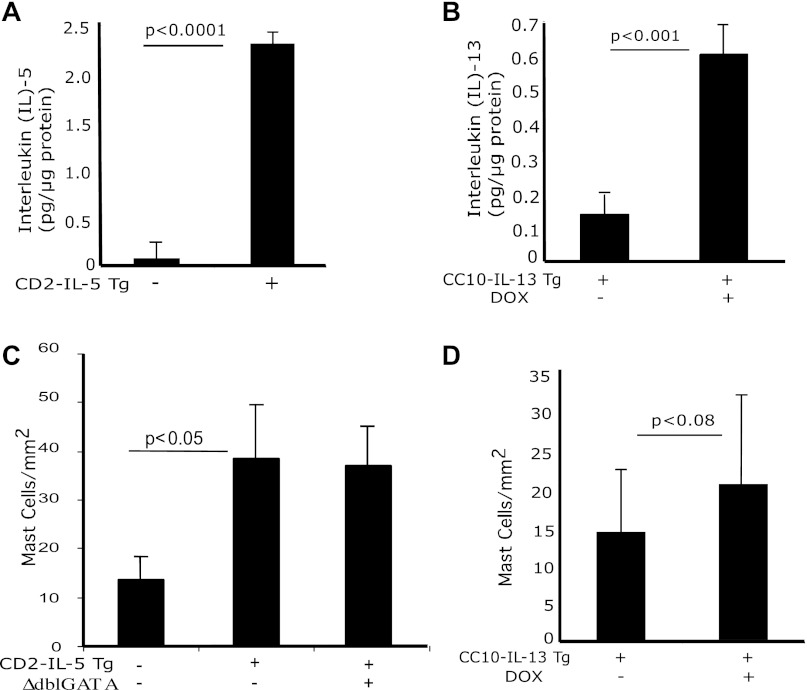
IL-5, IL-13, and mast cell induction in the esophagus. Protein levels of IL-5 and IL-13 from esophageal tissue of wild-type (WT), CD2-IL-5, and uninduced and doxycycline (DOX)-induced rtTA-CC10-IL-13 mice were measured by performing ELISA. Levels of IL-5 in 12-wk-old WT and in CD2-IL-5 mice and of IL-13 in 8-wk-old WT and in rtTA-CC-10-IL-13 mice are shown (A and B). The level of mast cells in the esophagus of WT, CD2-IL-5, ΔdblGATA/CD2-IL-5, and DOX- and no-DOX-treated rtTA-CC10-IL-13 mice were quantified by performing morphometric analysis following chloroacetate tissue staining (C and D). The results are the summary of 3 independent experiments, reported as means ± SE with n = 6 mice for each group. The statistical P values are provided in each figure.

