Fig. 3.
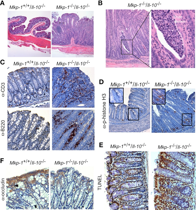
Mkp-1 deficiency exacerbates the epithelial proliferation and inflammation of the large intestine of Il-10 knockout mice. Mice were housed in a SPF environment and euthanized at 8 wk of age. The colon was excised and fixed for histology and immunohistochemistry analyses. The colon of wild-type (Mkp-1+/+/Il-10+/+) and Mkp-1 knockout (Mkp-1−/−/Il-10+/+) mice appeared normal (data not shown). A: histology of hematoxylin and eosin (H&E)-stained colon sections. Note the marked mucosal epithelial hyperplasia in the double knockout (Mkp-1−/−/Il-10−/−) section, but not in the Mkp-1+/+/Il-10−/− section. B: mucosal leukocytes infiltration with crypt abscess in the Mkp-1+/+/Il-10−/− section. Image on the right is a high-magnification image of the crypt abscess. C: immunohistochemical detection of T lymphocytes (anti-CD3, top) and B lymphocytes (anti-B220, bottom) in the colon section of the Mkp-1+/+/Il-10−/− and Mkp-1−/−/Il-10−/− mice. D: immunohistochemical detection of the mitotic marker phospho-histone H3 in the rectal mucosa of Mkp-1+/+/Il-10−/− and Mkp-1−/−/Il-10−/− mice. E: detection of apoptosis in the colon section of the Mkp-1+/+/Il-10−/− and Mkp-1−/−/Il-10−/− mice by TUNEL assay. F: immunohistochemical detection of α-occludin in colon, with enhanced positive (brown) signal in the Mkp-1+/+/Il-10−/− section than in the Mkp-1−/−/Il-10−/− section. After immunohistochemical staining the sections were counterstained with hematoxylin. Images shown are representative of at least 3 different sections.

